Meet Sherlock Holmes

Sherlock Holmes: Britain's best detective and the brilliant mind behind many of Scotland Yard's cases. His incredible deduction skills and extremely wide range of knowledge will surely blow you away- at 221B Baker Street, his home and workplace, Sherlock will guide you through the entire nervous system, one neuron by neuron.
Check It Out- Sherlock's Daily Routine
Under The Scope
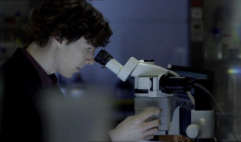
Sherlock is constantly at St. Bart's- you can find him at the science lab most of the time. Molly will usually be there too, helping Sherlock with his experiments and quite often, bringing his coffee. Take a look at what's under his microscope on the right side of the page. |

The Neuron
This is what Sherlock sees underneath the microscope: a neuron. It may not look like much, however, it is something that is essential to our body functions. You see, but not observe. The nervous system consists of the brain, the spinal cord, sensory organs, and these neurons. This system controls and coordinates our body functions. Neurons send and receive messages from one body part to another, thus neurons play an especially vital role. Neurons are masses of nerve cells that transmit information all over your body through nerve impulses, electrical signals that carries the message to your body. They consist of three main parts. The axon, the dendrite, and the cell body. Thin-branch like structures called dendrites protruding from the cell body pick up stimuli from the surrounding environment, other neurons, and sensory receptor cells. Axons also extend from the cell body to send signals onward to other neurons through neurotransmitters, chemicals released by one neuron to stimulate the neighbouring one. These neurotransmitters are released into the synapse which is a junction between one neuron and another. Let's take for example- acetylcholine, the most common neurotransmitter, stimulates muscle contractions in our body. Acetycholine is released from the axon terminal across the synapse to another neuron in a response to an electrical impulse. It's quite simple, isn't it? You're already prepared to discuss the nervous system with Sherlock- He'll be impressed. |
Sherlock's Mind Palace

When Sherlock is in need of information, he automatically goes into his "mind palace", which is his tool for remembering the smallest of details and organising his information in a visual way. His palace is essential when it comes to solving crimes- wonder how he manages to store all that information?
Let's take a look at the parts of his brain that allow him to do such extraordinary things:
Let's take a look at the parts of his brain that allow him to do such extraordinary things:
Sherlock's Extraordinary, Yet Ordinary Brain

On the right side is an image of the control centers of the brain. There are five main parts to this structure:
The Frontal lobe: associated with reasoning, planning, parts of speech, movement, problem-solving, and emotions.
The Parietal Lobe: also associated with movement, orientation, recognition, and perception of stimuli.
The Occipital Lobe: associated with visual processing
The Temporal Lobe: associated with perception, auditory stimuli, and speech.
The Cerebellum: Also called the "little brain" is associated with coordination of movement, posture, and balance.
Although the brain is the main control center of the body, the spinal cord is also an essential part of the nervous system. Together, they're called the Central Nervous System (CNS), where information is evaluated and decisions are made, sending a signal through the Peripheral Nervous System (PNS) which are the nerves of the body.
Look down below to see an organised tree chart of the different branches of the nervous system.

The Peripheral Nervous System is divided into two parts: sensory neurons and motor neurons.
Sensory neurons typically have a long dendrite with a short axon, receiving and carrying information from the sensory receptor cells to the Central Nervous System.
On the other hand, Motor neurons tend to have a long axon with a short dendrite and transmit messages from the CNS to the muscles and glands.
These motor neurons are also classified into two parts, the somatic nervous system and the autonomic nervous system. The somatic nervous system is in charge of voluntary movement, mostly referring to the skeletal muscle, whereas the autonomic system is in charge of involuntary movement in charge of cardiac muscles, smooth muscles, and glands.
The autonomic system is subdivided into two parts- the sympathetic and parasympathetic. The sympathetic system is prepares the body to take action, for example, in a dangerous situation by increasing the heart rate, thus called "fight or flight" responses. However, parasympathetic does quite the opposite. In charge of the "rest and digest" responses, it activates tranquil functions, such as conserving energy by slowing the heart rate and stimulating the secretion of saliva or enzymes into the stomach.
A Free Meal, You Say?
Have a free dinner at Angelo's- courtesy of Sherlock Holmes. A couple years ago, Sherlock cleared Angelo's name as a suspect in two brutal murders when Lestrade and the police thought he had committed the crime. Sherlock proved that Angelo was on the other side of London stealing a car at the time of the murder, which cleared his name (only slightly) as he was arrested for robbery. Angelo is always overjoyed to see Sherlock at his restaurant, giving him free meals to show his thanks. He is more than willing to serve Sherlock and his acquaintances!
|
Caution!
Judging by the dangerous, action-packed lives of Sherlock and John, surely the sympathetic nervous system is extremely important when it comes to solving their cases!
At Baker Street, be sure to prepare yourself for what you might find. Sherlock tends to have his experiments set out everywhere from the living room to the kitchen- so be very careful. This package is not recommended to anyone who may not be able to handle gruesome sights; Sherlock is known for keeping his experiments out and around in his flat. Ranging from eyeballs in the microwave to decapitated heads in the fridge, it is a rather shocking sight, not recommended for the faint-hearted. |
